With cone beam computed tomography, oral health professionals gain a highly accurate 3-D image of the patient’s anatomy from a single scan. These 3-D images allow the practitioner to better diagnose and understand the true extent of dental disease, and they can provide for more appropriate treatment for patients.
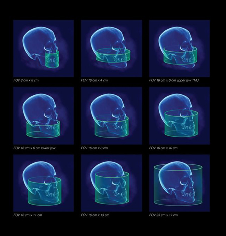
- FOV 16cm x 4cm : Single Dental Arch
- FOV 8cm x 8cm : Both Dental Arches
- FOV 16cm x 6cm : Maxilla/Mandible
- FOV 16cm x 8cm : Maxilla and mandible
- FOV 16cm x 10cm : Maxilla, mandible and TMJ
- FOV 16cm x 11cm : Maxilla, mandible, TMJ and ENT
- FOV 16cm x 13cm : Maxillofacial including orbits
- FOV 23cm x 17cm : Cranofacial
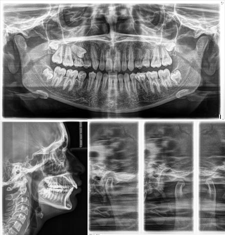
- Panoramic radiograph
- Bitewing panoramic radiographs
- TMJ Open and Closed Lateral, PA and Cross-sectional views
- Maxillary sinus view
- Lateral cephalogram
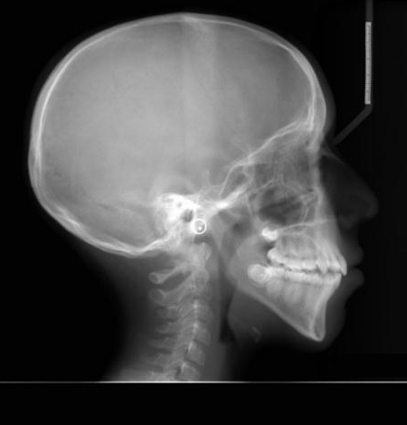
- Lateral skull view
- Hand-wrist radiograph
- Extra-oral skull radiography
IOPA, bitewing, occlusal - coming soon
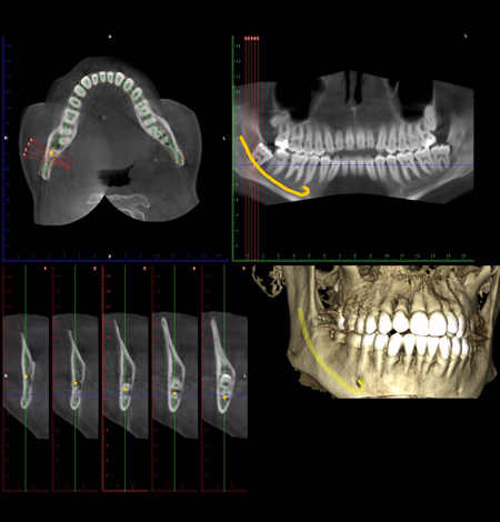
- Pathology diagnosis and localization
- Implant site assessment and virtual simulation
- 3D Cephalometric analysis
- Airway and Paranasal Sinuses analysis
- TMJ osseus pathology assessment
- Orthognathic surgery workup
- 3D study model design and printing
* We also provide on-site diagnostic and analytic services, on an appointment basis.
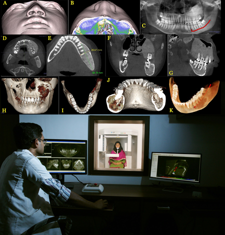
- Online radiology interpretation and reporting services for images taken from other centres
- Assist students and researchers in medical and dental imaging applications
- Conduct continuing educational programmes for clinicans and postgraduates
- Hands-on workshops and software training on various applications of CBCT
- Maintain a database of rare cases for future references